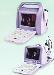
Full Digital Ophthalmic Ultrasound Scanner

Full Digital Ophthalmic Ultrasound Scanner
Full Digital Ophthalmic Ultrasound Scanner
Keeping sync with latest industrial demands, we are involved in manufacturing, supplying and trading highly effective Full Digital Ophthalmic Ultrasound Scanner. Our offered scanners are very much popular in the market for their precise functionality, user-friendly controls and non-radioactive attributes. These scanners are designed and fabricated in various stipulations by using high-grade raw materials and progressive technology. Apart from this, we intend to supply our range of products at competitive market rates.
Features:
High accuracy
Easy to use
Reliable operation
Full Digital Ophthalmic Ultrasound Scanner A/B
Perfect combination of A scan and B scan: A scan measures the distances by forming single dimension image, e.g. ACD, lens, vitreous, AXL', eye axis related diseases etc. B scan converts the tissue echos into echo luminous spots with different brightness by sector scanning, which symbolize the intensity of echos. The clear and intuitive image helps to observe the intra-ocular and posterior segment diseases. Real-time dynamic scanning shows the lesion locations, volume, shape and relations with peripheral tissues. B scan has been widely used in the diagnosis of ocular and orbit diseases, including:
Turbid refractive media: ultrasound scanning is the best option to show the intra-ocular diseases.
Somatometry of the eye
Diagnosis before and after vitrectomy to confirm the range and extent of disease.
Diagnosis and location of ocular trauma and intraocular foreign body.
Intra-ocular tumor
Diagnosis of exophthalmos
Ocular and orbit hemodynamics
Intrusive ultrasound to take out non-magnetic intraocular foreign body; tumor puncture under ultrasound guidance.
A- Scan:
Display: B, B/B, 4B , B/A, A five-point measure, A single point measure
Measure Range: A Five-point: 0~36mm; A single-point: 0~72mm
Measure: ACD, Lens, Vitreous, CCT, AXL(immersion mode)
Eye type: Normal/Aphakic/ Dense Catract/ Manual
Measure mode: Immersion and contact, manual and auto measurement
Calculation : std. Deviation, Average, auto average measurement of eight
groups , result correction,
Measurement method: A five-point measurement, A single-point measurement IOL formulas: SRK-II, SRK-T, BINKHORST, HOLLADAY, HOFFER-Q, HAIGIS,
Display of random two formulas, calculation of degree of IOL, DEM of
emmetropization of IOL, DAM of IOL, REEF after surgery,
100 groups of A scan results storage
A single point measurement:
Magnification: x1.0, x2.0, x4.0
Depth display: 0~36mm
Delayed depth: 0~36mm
B-Scan:
Scan mode : sector scanning, stepping motor with high precision
Comparison of two or four images
Depth selection: 24mm, 31 mm, 38mm, 42mm, 45mm,
49mm, 52mm, 56mm total 8 grades
Compression Curve:8
Post-processing:8
Postponed depth range: 25-32mm Local zoom Up/down, L/R image tuning Color option:8
Image storage :more than 10,000 images
Cine loop:256 frames, measurement available
Annotation: input character and arrow, Chinese/English available
Disk management
Measurement: distance, circumference, volume, angle, histogram, sectional view, etc.
Report print: screen copy, IOL report, medical record with image etc. Language :Chinese/English
USB2.0 to support U disk(file manipulation, software upgrade, one -key storage, etc)
Internal D disk(4G) for data, image , report, movie clip
Send Enquiry

